PUBLICATIONS
DIAMONDVIEW™, CONCEPT AND USE AT LFG
Aurélien DELAUNAY
The French Gemmology Laboratory has been equipped with a latest-generation DiamondView since 2006 (Figure 1). The DiamondView™ is a low wavelength luminescence imaging device (approximately 225 nm) developed by the DTC (De Beers) in 1996. Its high energy ultraviolet light source makes it possible to observe the sometimes complex growth morphology of diamonds (Figure 2).
Figure 1: DiamondView™ used at LFG
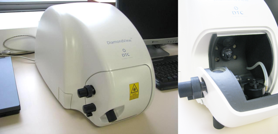
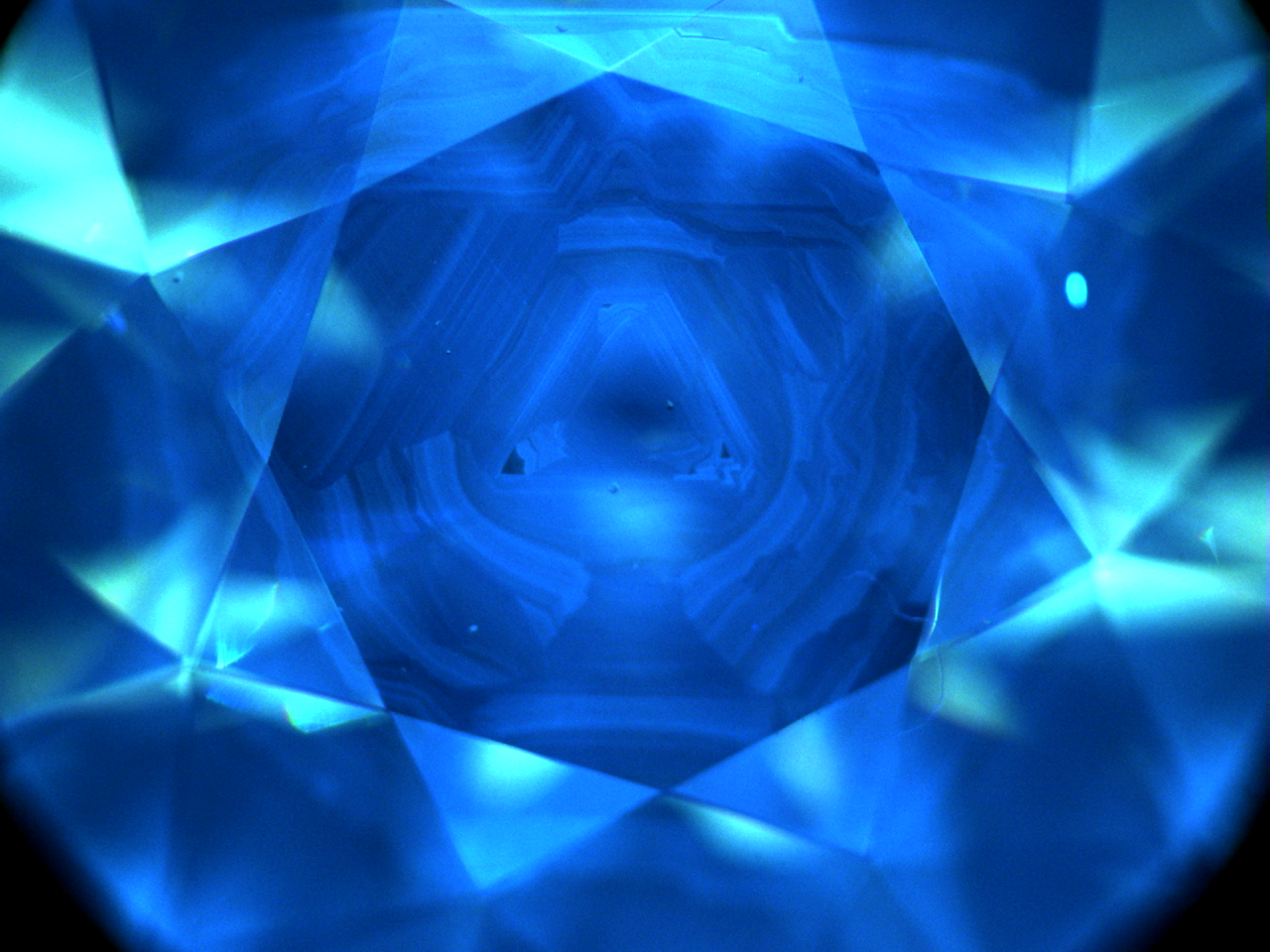
Figure 4: infra-red spectrum of the J18 diamond previously observed under UV, typical of a pure Ib HPHT synthetic diamond. The isolated nitrogen atoms, or C centre, are characterised by absorptions at 1045, 1130, 1344 and 2688 cm(1).
The potentially synthetic diamonds isolated by their luminescence and/or infra-red spectrum are then observed under the microscope. Typical inclusions such as scattered pinpoints (), inclusions in “bread crumb” formation can be characteristic of HPHT synthetic diamonds (Figure 5a and 5b).
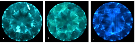
Figure 3: DiamondView images of CVD synthetic diamond (a), HP-HT synthetic diamond (b), and natural diamond (c)
Moreover, a diamond’s growth morphology is an exact reflection of its DNA and cannot be modified during recut. If a diamond is returned to the lab or another lab, the DiamondView™ luminescence images are good ways to recognize it. Today, the LFG records DiamondView™ images for all isolated diamonds analysed.
DiamondView™ is also helpful when analysing other gems such as corundum. The luminescent growth sectors, their position and colour offer us valuable insights into gem analysis. DiamondView™ images can point to a certain geographic origin or specific form of treatment.
It is therefore a “precious” device for the laboratory in the analysis of diamonds, and whose applications and developments to other gems remain to be invented!

