MICRORADIOGRAPHY – TOMOGRAPHY
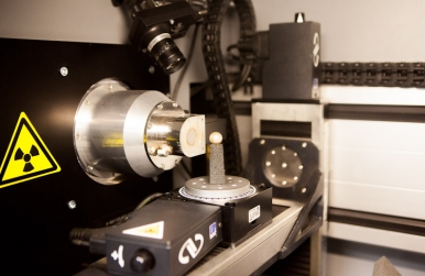
This X-ray imaging technique makes it possible to visualise the internal structures of pearls and therefore to distinguish fine pearls from cultured pearls. It also makes it possible to distinguish cultured beads with core from cultured pearls without core (keshi).
The combination of tomography with microradiography technology makes it possible to reconstruct the volume studied in 3D (in this case, the pearl) and therefore makes it possible to view precise cuts in all directions of space. This tool has proved in analysing pearls, and in particular discerning keshi (pearls of cultures without core) from fine pearls.
The camera also makes it possible to visualise the luminescence of the pearls analysed on x-rays to define their growth environment: fresh water or seawater.
CONTACT US :
contact@lfg.paris
(+33) 01 40 26 25 45
30, rue de la Victoire - 75009 Paris
Monday - Thusday : 9H30 to 17H00
Friday : 9h30 to 16h30
Withdrawal files at same days and time